


Clinical Cognitive Neuroscience Center
Research
Neuroimaging
We combine multiple neuroimaging modalities to study brain structure, connectivity, function, and molecular processes in psychiatric disorders.
Magnetic Resonance Imaging (MRI)
Brain Structure, Connectivity, and Function
Structural MRI
We measure cortical thickness, gyrification, and subcortical volumes, and apply MRI texture analysis to capture microstructural alterations that conventional morphometric measures may miss. We also use neuromelanin-sensitive MRI as an indirect marker of dopamine function, and apply hippocampal surface modeling in collaboration with Ilwoo Lyu (POSTECH).
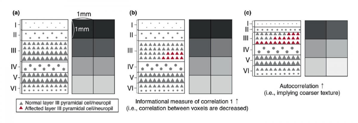
Schematic illustrating how microstructural changes in cortical layers may alter MRI texture features (Moon et al., Mol Psychiatry, 2023).
Diffusion MRI
We use diffusion tensor imaging (DTI) with probabilistic tractography to map white matter connections between brain regions, and diffusion kurtosis imaging (DKI) to assess tissue microstructure beyond conventional DTI measures.
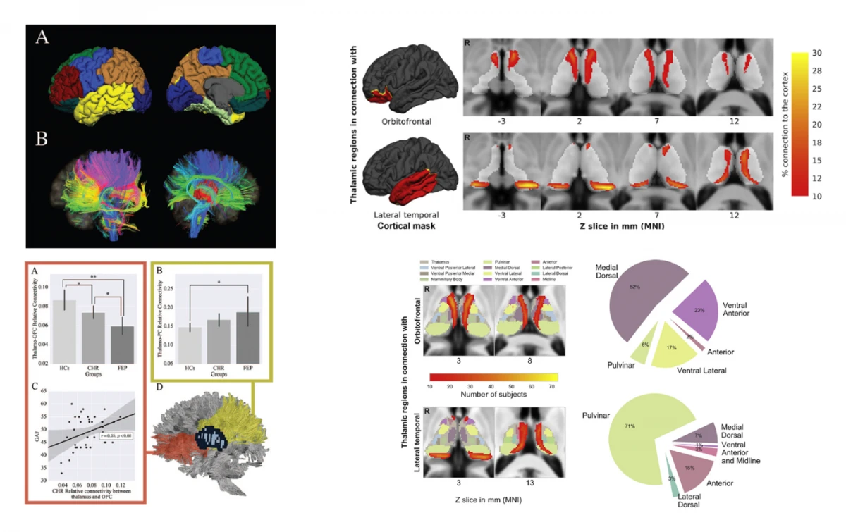
Left: probabilistic tractography of thalamo-cortical white matter connectivity in individuals at clinical high risk and with first-episode psychosis (Cho et al., Schizophr Bull, 2016). Right: connectivity-based segmentation of the thalamus combined with DKI to examine the microstructure of higher-order thalamic nuclei (Cho et al., Biol Psychiatry, 2019).
Functional MRI
We examine resting-state functional connectivity within and between large-scale brain networks, using parcellations of the cortex, thalamus, and cerebellum to study circuit-level interactions.
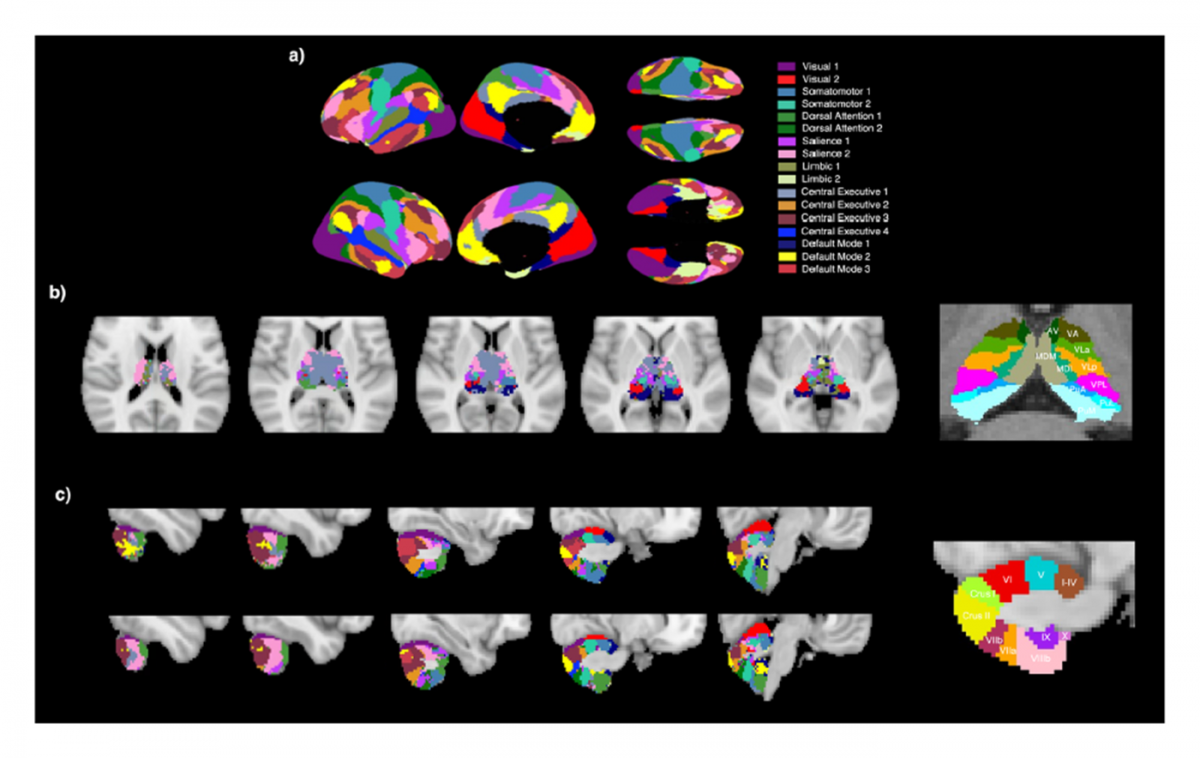
Cortical network, thalamic, and cerebellar parcellations used to model cortico-thalamo-cerebellar network interactions (Ha et al., Schizophrenia, 2023).
Positron Emission Tomography (PET)
Molecular Imaging of the Living Brain
We use PET to measure molecular processes in the living brain, including reactive astrocytes with [18F]THK5351 and dopamine D2/3 receptor occupancy by antipsychotic drugs.
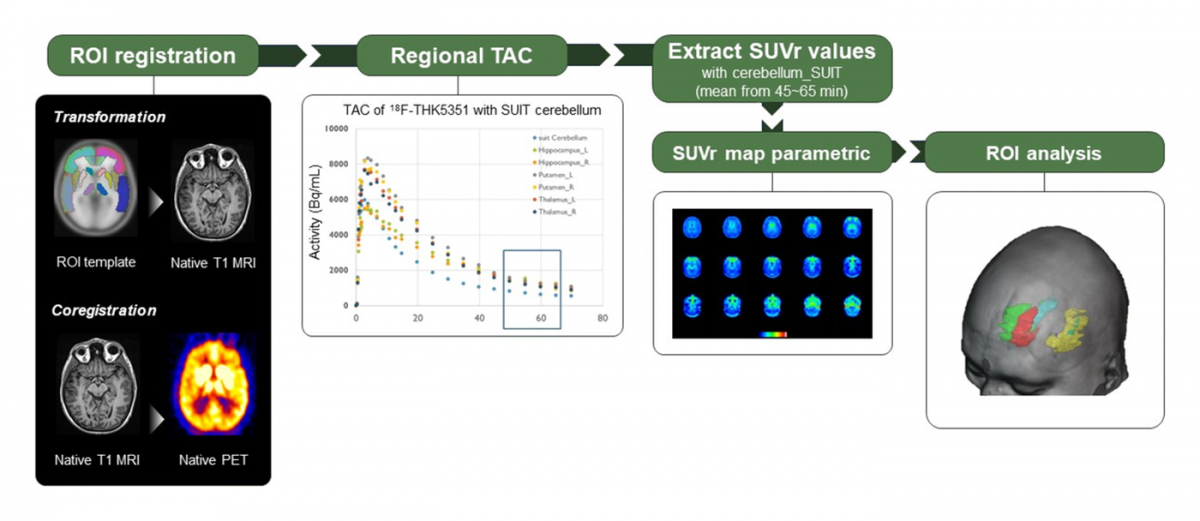
Analysis pipeline for [18F]THK5351 PET, from region-of-interest registration and regional time–activity curves to standardized uptake value ratio (SUVR) mapping and region-of-interest analysis (Kim et al., JAMA Netw Open, 2024).
Computational Modeling of Brain Stimulation
Toward Personalized Neuromodulation
Building on our multimodal imaging data, we are developing personalized models of brain stimulation that combine individual electric field simulations with whole-brain network models fitted to each person’s structural and functional connectivity.
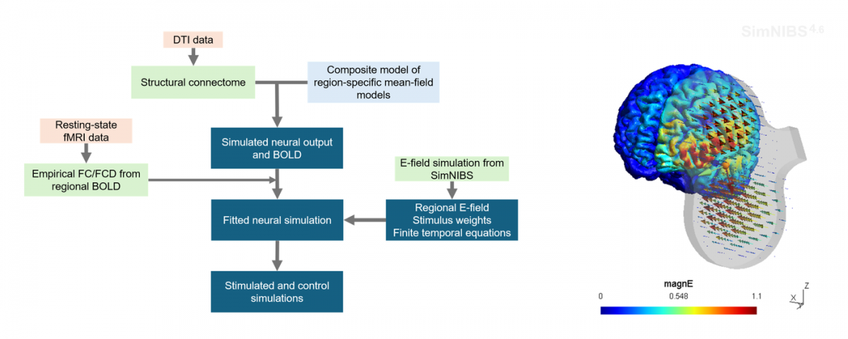
Pipeline for personalized whole-brain simulation of brain stimulation, integrating DTI-based structural connectomes, resting-state fMRI, and individual electric field simulations (right: an example electric field model generated with SimNIBS).